Skip to content
High-resolution Endoscopy with NBI, BLI and LCI Imaging

Procedure

High-resolution Endoscopy with NBI, BLI and LCI Imaging

Modern endoscopes combine high-resolution video with specialised light filters (NBI, BLI, LCI) that highlight surface vessels and tissue patterns, helping us detect very early changes.

What is it?

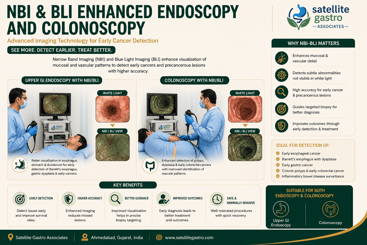
Narrow Band Imaging (NBI), Blue Laser Imaging (BLI) and Linked Colour Imaging (LCI) use light filters to enhance the visibility of mucosal patterns and blood vessels.

These features come built into our endoscopes and are used during routine procedures for any patient where they add value.

Who needs it?

  • Surveillance of known Barrett oesophagus.
  • Surveillance after gastric or colonic polyp or cancer removal.
  • Detection of early changes in long-standing reflux or H. pylori-related gastritis.

How it works

No different from standard endoscopy. The advanced imaging modes are activated with a button on the scope handle as needed.

Preparation

  • Same as standard endoscopy.

Recovery

  • Same as standard endoscopy.

Risks

All endoscopic procedures carry a small risk of bleeding, perforation, or reaction to sedation. These risks are uncommon when the procedure is done by a trained endoscopist in a hospital setting.

We will explain the risks specific to your case during the consultation, before you sign consent.

Common questions

Things patients ask us.

Is this the same as a regular endoscopy?

Same procedure, better cameras. The enhanced imaging modes are used selectively when they add information.

Related procedures

Conditions we treat with this

Next step

Speak with a consultant about High-resolution Endoscopy (NBI / BLI / LCI).

Locale: hi